A luxated lens can seriously impact your dog’s vision. Early detection is crucial. Many pet owners don’t recognize the signs until it’s too late. This condition affects thousands of dogs yearly. The lens plays a vital role in focusing light onto the retina.
When it becomes displaced, vision problems occur rapidly. Dogs of all ages can develop this condition. Certain breeds face higher risks. Understanding treatment options is essential for making informed decisions. The cost of treatment varies widely depending on location and severity. Your swift action could save your dog’s eyesight.
Modern veterinary medicine offers several effective treatments. Each case requires individual assessment. The emotional impact on pets and owners shouldn’t be underestimated. Regular eye checks can catch problems early. This comprehensive guide covers everything you need to know about lens luxation in dogs.
Understanding Luxated Lens in dogs
The canine eye contains a lens that focuses light. This lens sits behind the pupil. It’s normally held in place by tiny fibers. These fibers are called zonules. They anchor the lens to the ciliary body. When these fibers break or weaken, the lens can shift position.

This displacement is called lens luxation. It’s a serious condition requiring immediate attention. The displaced lens can cause increased pressure within the eye. Without treatment, permanent vision loss may occur. The condition can be extremely painful for dogs. You might notice your dog pawing at their eye.
Some dogs may become withdrawn due to discomfort. Hereditary conditions often trigger this problem in certain breeds. Early detection gives your dog the best chance for successful treatment. Vision preservation depends on quick intervention. Many dogs with this condition show obvious signs of discomfort.
There are two main types of lens luxation:
Different types of luxation require different treatments. Understanding the specific type affecting your dog is crucial. Veterinarians classify luxation based on the direction of lens movement.
Each type carries its own set of risks and complications. Treatment success rates vary between types. Your vet will determine which type your dog has through careful examination. This diagnosis guides the treatment approach.
1. Anterior Lens Luxation
In anterior luxation, the lens moves forward. It shifts into the front chamber of the eye. This type is more dangerous. It often causes immediate pressure increases.
The displaced lens blocks normal fluid drainage. This blockage can quickly lead to glaucoma. Pain develops rapidly with this type. The eye may appear cloudy or red. Vision loss can happen within hours or days. Emergency treatment is absolutely necessary.
Surgical intervention is usually required. Delays in treatment can result in permanent blindness. This type occurs more frequently in dogs. Many veterinarians consider this type a true emergency. The prognosis depends on how quickly treatment begins.
2. Posterior Lens Luxation:
With posterior luxation, the lens moves backward. It shifts toward the back of the eye. This type is generally less urgent. It typically causes fewer immediate complications. The lens displaces into the vitreous chamber. Pain may be less obvious with this type.
Vision impairment still occurs but develops more gradually. The eye might still look relatively normal. Your dog may adapt to vision changes more readily. Posterior luxation can sometimes be managed without surgery.
Regular monitoring becomes essential. This type can eventually progress to anterior luxation. Some dogs with mild cases maintain functional vision. Your vet will advise on the best approach for your pet’s specific situation.
Causes and Risk Factors
Genetic mutations play a significant role in lens luxation. Many cases have a hereditary component. Certain dog breeds face much higher risks. Terriers top the list of predisposed breeds. Jack Russell Terriers show particularly high rates.
Border Collies also face elevated risks. Shar Peis and Chinese Crested dogs are vulnerable too. The condition typically appears in middle-aged dogs. The hereditary form usually emerges between ages 3-8. Some cases result from trauma to the eye. Injuries can damage the supporting fibers. Chronic eye inflammation increases risk factors.
Secondary luxation follows other eye diseases. Glaucoma commonly precedes lens luxation. Advanced cataracts may contribute to the condition. Age-related degeneration weakens supporting structures. Diabetes can increase vulnerability to eye problems. Previous eye surgeries sometimes lead to complications. Understanding your dog’s specific risk factors helps with prevention. Regular screenings are vital for high-risk breeds.
Symptoms to Watch Out For
Early detection saves vision. Watch for these warning signs. Your dog may squint frequently. Excessive tearing often occurs. The affected eye might appear cloudy. Red or bloodshot eyes signal trouble. Increased pawing at the eye suggests pain.
The eye might look bluish or hazy. You may notice the pupil appears irregular. Some dogs develop obvious discomfort in bright light. Changes in eye color can indicate problems. The white of the eye may look inflamed. Your dog might bump into objects. Behavioral changes often accompany eye pain. Sudden reluctance to play could signal issues.
Loss of appetite may develop with serious pain. Head tilting sometimes indicates eye discomfort. Vision changes may cause startled reactions. Cloudy fluid might be visible within the eye. Discharge from the eye requires attention. Compare both eyes for subtle differences. Any sudden changes warrant immediate veterinary care.
Diagnosis and Treatment
Veterinary ophthalmologists specialize in eye conditions. They use special equipment for thorough examination. Diagnosis begins with a complete eye exam. The vet will check intraocular pressure. They’ll examine the position of the lens capsule.
Dilating drops help visualize internal structures. The vet may use an ophthalmoscope. This tool allows detailed examination of eye structures. Ultrasound imaging might be necessary in some cases. Blood tests rule out underlying conditions. Treatment depends on the type of luxation. The degree of displacement affects treatment options.
The presence of other eye diseases matters significantly. The vet will assess your dog’s overall health. Age and breed influence treatment decisions. The condition of the affected eye guides intervention choices. Vision prognosis varies considerably between cases. Early diagnosis substantially improves outcomes. Your vet will explain all suitable options.
Ty’s Journey: Navigating Lens Luxation in His Senior Years
Ty was a 13-year-old Maltipoo. His owner noticed something wrong with his eye. The initial diagnosis was a simple dry eye. Treatment began with standard eye medications. Unfortunately, his condition worsened rapidly. The lens had completely dislocated.
Pressure increased dangerously in his eye. Emergency intervention became necessary. Ty underwent surgery to save his vision. Despite efforts, his eye couldn’t be saved. Removal became the only option to relieve pain. The surgery went smoothly without complications.
Ty adapted remarkably well to having one eye. His quality of life improved once the pain subsided. His owner now carefully monitors his remaining eye. Early intervention would be possible if signs appear in the other eye. Ty’s case demonstrates the importance of prompt attention. Senior dogs may have additional surgical risks. Ty’s journey highlights the emotional aspects of pet health decisions.
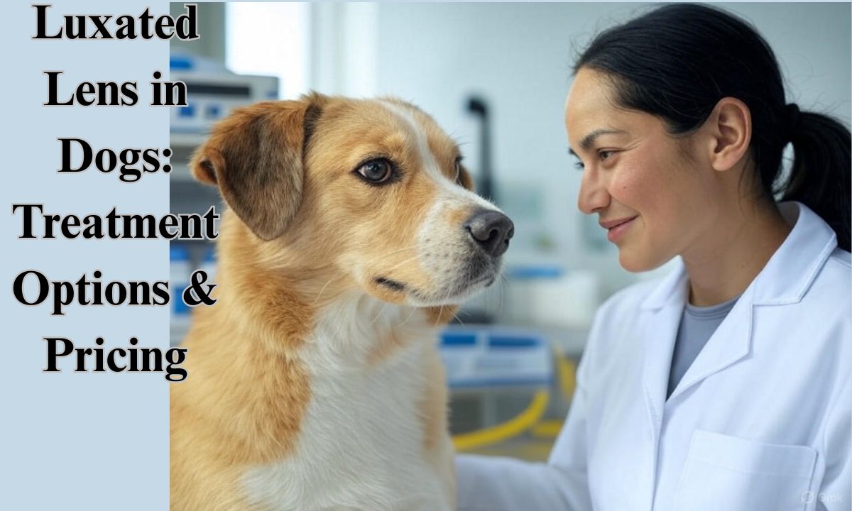
Treatment Options & Cost
Treatment options vary based on several factors. Surgical intervention is often necessary. Medication alone rarely resolves the condition. Costs vary widely by location and severity. Basic lens removal typically costs $1,500-$3,500. Specialized surgical techniques may cost more.
Enucleation (eye removal) ranges from $1,000-$2,000. Emergency treatment adds to overall costs. Metropolitan areas generally charge higher fees. Specialist referral centers have premium pricing. Some pet insurance policies cover these procedures.
Post-operative medications add additional expenses. Follow-up appointments are necessary for monitoring. Pain management medications will be needed. Protective collars prevent self-injury during recovery. Some cases require lifelong medication.
Rural areas may offer more affordable options. Many clinics offer payment plans for expensive procedures. The cost of inaction usually exceeds treatment costs. Vision preservation justifies the investment for most owners. The specific surgical approach influences overall pricing.
Ty’s Difficult Choices: Navigating the Options for Lens Luxation
Ty’s owner faced difficult decisions. Three main treatment options were presented. Each carried different risks and costs. Lens couching was the first option presented. This procedure would push the lens backward. Success rates hover around 50%.
The cost estimate was $3,500. Complete lens removal was the second option. This would preserve the eye’s appearance. Lifelong medication would be required afterward. This option carried a $7,000 price tag. Complete eye removal was the final option. This would permanently eliminate pain. The cost was approximately $4,000.
Ty’s owner carefully weighed each option. The decision process involved significant emotional stress. The veterinarian provided detailed information about each approach. Quality of life remained the primary consideration. The success rates influenced the final decision. Ty’s owner chose lens couching initially. Unfortunately, this procedure proved unsuccessful. Eye removal became necessary the following day. The experience demonstrated the unpredictable nature of eye surgeries.
Lens Luxation in Cats
Cats can also develop lens luxation. The condition presents similarly to dogs. Trauma causes more cases in cats than in dogs. Feline cases often result from injuries. Cats show different clinical signs than dogs. They may hide symptoms more effectively.
Diagnosis follows similar procedures as with dogs. Many treatment options remain the same. Surgical approaches require slight modifications for cats. The prognosis varies depending on cause and timing. Emergency intervention is equally important for cats. Some breeds show higher predisposition. Persian cats face elevated risks.
Cost ranges remain comparable to canine procedures. Recovery protocols differ slightly for felines. Cats generally tolerate eye removal well. Adaptation to vision changes occurs quickly in most cats. The behavioral impact may be less obvious. Early detection remains crucial for successful outcomes.
READ THIS BLOG : Unraveling the Spiritual Meaning of a Black Cat Staring At You
Prevention and Aftercare
Prevention begins with knowing your dog’s risk factors. High-risk breeds need regular eye examinations. Annual checkups should include thorough eye assessment. Early intervention prevents serious complications. Aftercare following surgery is critical.
Follow all medication schedules precisely. Restrict your dog’s activity during recovery. Prevent rubbing or scratching of the surgical site. Protective collars are essential during healing. Watch for signs of infection or complications. Keep follow-up appointments without fail. Clean the area as directed by your veterinarian.
Some dogs require special housing arrangements during recovery. Maintain a calm environment to reduce stress. Monitor food and water intake during recovery. Check the surgical site daily for changes. Report any concerns to your veterinarian immediately. Complete the full course of prescribed antibiotics. Pain management improves recovery outcomes. Consider environmental modifications for vision-impaired pets.
Frequently Asked Questions
What is lens luxation?
Lens luxation occurs when the eye’s lens displaces from its normal position. The lens moves either forward or backward within the eye. This happens when the fibers holding it in place break or weaken. The condition causes pain and vision problems without treatment.
What causes lens luxation in dogs and cats?
Most cases stem from hereditary conditions affecting certain breeds. Terriers face particularly high risks due to genetic factors. Trauma can displace the lens in some cases. Other eye diseases like glaucoma sometimes lead to secondary luxation.
If one eye has a luxating lens, does it mean the other one will too?
Not necessarily, but the risk is substantially higher. Genetic cases often eventually affect both eyes. The second eye requires careful monitoring. Early intervention can prevent complications if signs appear.
How can I tell if my pet has lens luxation?
Watch for squinting, redness, or cloudiness in the eye. Excessive tearing often occurs. Your dog may paw at the affected eye. Vision changes might cause bumping into objects. Any sudden eye changes warrant immediate veterinary attention.
What are the treatment options for lens luxation?
Treatment depends on luxation type and severity. Surgical lens removal relieves pressure and pain. Eye removal becomes necessary in advanced cases. Some posterior luxations may be monitored without immediate surgery. Medication helps manage symptoms but rarely resolves the
Conclusion
Lens luxation presents serious challenges for dogs and their owners. Understanding the condition is the first step toward proper management. Early detection dramatically improves outcomes. Know your dog’s risk factors based on breed and age. Regular veterinary examinations help catch problems early. The condition requires prompt professional attention. Treatment options have improved significantly in recent years.
The financial investment protects your dog’s quality of life. Work closely with your veterinarian to create a treatment plan. Each case requires individualized care approaches. The prognosis depends largely on how quickly treatment begins. Many dogs adapt remarkably well after appropriate intervention. Vision preservation remains possible with timely care. Even complete eye removal can restore comfort and quality of life. This condition reinforces the importance of routine eye examinations. Your vigilance as an owner plays a crucial role in your dog’s eye health.

jack is an experienced blogger and a passionate wordsmith at Phrase Pioneers. With a keen eye for language and a deep love for writing, she shares insightful posts on grammar, phrases, and the art of communication.